




Emergency 0484 7185000

 Second Opinion
  Book
an Appointment
Book
an Appointment
 Our Doctors
Offcanvas
Emergency
0484 7185000
Call Us
8593882299
Centres of Excellence




0484 7185000
8593882299
Accurate diagnosis is the foundation of effective medical treatment. Advanced imaging technologies allow doctors to examine internal organs, blood vessels, bones and soft tissues in detail, often before a condition produces serious complications.
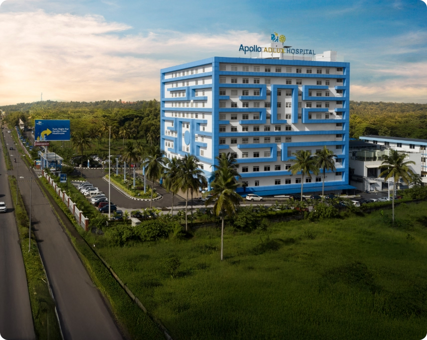
Apollo Adlux Hospital, Kochi, is equipped with advanced diagnostic imaging facilities, including a 3 Tesla MRI scanner and a 128-Slice CT scanner. These technologies support the timely evaluation of neurological, cardiac, orthopaedic, abdominal, oncological and emergency conditions.
Magnetic Resonance Imaging, or MRI, uses a powerful magnetic field and radiofrequency signals to create detailed images of the body. The term “3 Tesla” refers to the strength of the MRI scanner’s magnetic field.
A 3 Tesla MRI scanner can produce high-resolution images that help doctors study complex anatomical structures, including the brain, spine, joints, nerves, muscles, blood vessels and internal organs.
Unlike CT scans and conventional X-rays, MRI does not use ionising radiation.
3 Tesla MRI may be recommended for the evaluation of:
The high field strength can support detailed anatomical assessment, although the most appropriate imaging protocol will depend on the patient’s symptoms and clinical condition.
Computed Tomography, commonly called a CT scan, uses X-rays and computer processing to create cross-sectional images of the body. A 128-Slice CT scanner acquires multiple image slices during a single rotation, enabling doctors to examine internal structures quickly and in detail.
CT imaging is especially valuable in medical emergencies because images can be obtained rapidly.
A 128-Slice CT scan may assist in diagnosing:
CT angiography may also be used to visualise blood vessels and identify narrowing, blockage, aneurysm or other vascular abnormalities.
3 Tesla MRI or 128-Slice CT: Which Scan Is Required?
MRI and CT scans serve different clinical purposes.
MRI generally offers excellent soft-tissue contrast and is frequently used for the brain, spine, nerves, muscles, ligaments and joints. CT scans are faster and are often preferred for trauma, fractures, lung conditions, internal bleeding and selected emergencies. The treating doctor or radiologist will recommend the most suitable scan after considering the patient’s symptoms, medical history and urgency of diagnosis.
Preparing for an MRI or CT Scan
Patients should inform the medical team about:
Some examinations may require fasting or the administration of contrast material. Patients will receive instructions based on the type of scan prescribed.
Advanced Diagnostic Imaging at Apollo Adlux Hospital, Kochi
Apollo Adlux Hospital brings together advanced radiology infrastructure and experienced medical professionals to support accurate diagnosis and coordinated treatment planning.
The availability of 3 Tesla MRI and 128-Slice CT Scan in Kochi strengthens the hospital’s ability to manage routine diagnostic requirements as well as neurological, cardiac, oncological and trauma-related emergencies.
Early and accurate imaging can help doctors identify the cause of symptoms, assess the extent of disease and determine an appropriate treatment pathway.